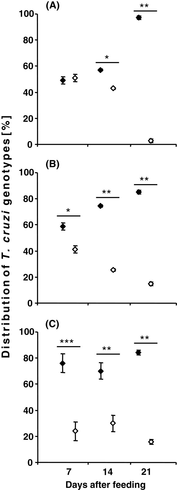
Figure 4.
Graphical illustration of the T. cruzi mini-exon amplicon intensity of mixed infections in different intestinal regions, small intestine (A), rectal lumen (B) and rectal wall (C) of R. prolixus mixed infections at daf, based on the results obtained by mini-exon PCR. Standard deviation is shown for each analysed sample. Statistically significant differences (* p < 0.05, ** p < 0.01, *** p < 0.001) are shown above the values. (♦ - C45/TcI, ◊ - JCA3/TcII).

