
Figure 2.
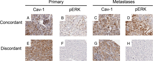
Concordance of Cav-1 and pERK-1/2 between matched primary and secondary. Typical immunohistochemical staining in matched primary and secondary tumours showing (i) positive concordance of both Cav-1 and pERK-1/2 (2A-2D) and (ii) discordance of Cav-1 and pERK-1/2 (2E-2H).