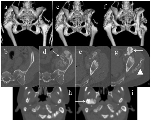
Figure 4.

CT images form Case 3. Reconstructed 3D-CT images of the hip joints at baseline (a), at M-12 m (c), and at D-12 m (f). Axial CT images of the left pelvis at baseline (b), at M-12 m (d, e), and at D-12 m (g), and those of the bilateral jaws at M-12 m (h), and at D-12 m (i). Massive new bone formations in the left iliac muscle were developed during the medication phase (d, arrow). During the discontinuation phase, the left intra-iliopsoas ossification matured (g, arrow), and heterotopic boned in the left gluteus medius (g, arrow head) and the right jaw joint (i, arrow) were newly formed.
