Figure 3.
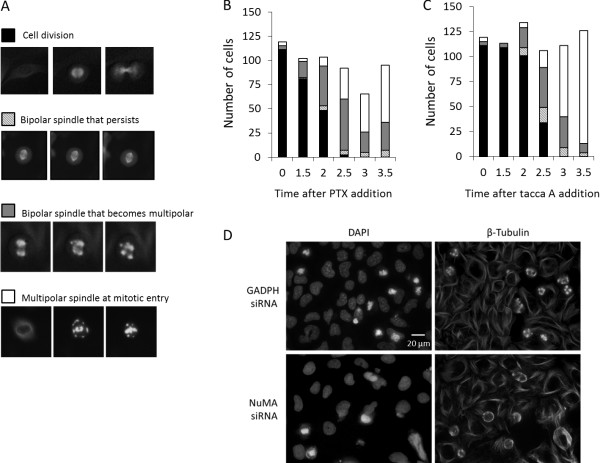
Aster formation following the addition of paclitaxel or taccalonolide A. GFP-β-tubulin expressing HeLa cells were treated with 12 nM paclitaxel or 5 μM taccalonolide A. Every cell that entered mitosis within 3.5 h after drug addition was followed until 8 h after drug addition and placed in one of 4 categories. (A) Representative images of the 4 categories of cells entering mitosis: bipolar spindle formation followed by cell division, bipolar spindle formation that persisted without completion of mitosis, bipolar spindle that resolved into multiple asters or formation of multiple asters immediately upon mitotic entry. Each image represents approximately 40 μm. The category of cells entering mitosis at the indicated time points following (B) paclitaxel or (C) taccalonolide A addition are shown. Cells in 60 individual microscopic fields from 12 separate wells were analyzed for each condition at each time point. (D) Effects of NuMA depletion on taccalonolide A-induced asters. Microtubules were visualized by indirect immunofluorescence 4 h after the addition of 5 μM taccalonolide A in control (GADPH siRNA) and NuMA-depleted (NuMA siRNA) HeLa cells (right). DNA was visualized by DAPI staining (left).

