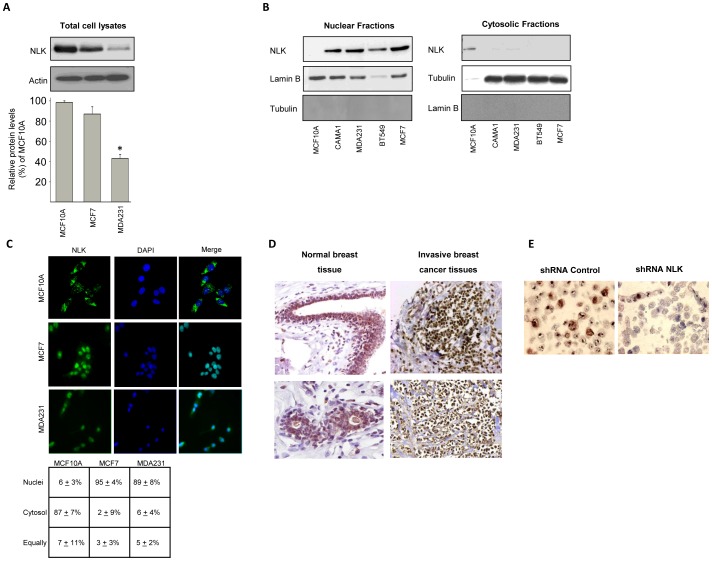
Figure 1. Endogenous NLK is localized in the nuclei of breast cancer cells and breast tumor tissue.
(A) The levels of NLK in total cell lysates of MCF10A, MCF7, and MDA231 (upper panel). Densitometric analyses of the total levels of NLK in MCF7, and MDA231 cell lines (Lower panel) relative to NLK protein expression levels in MCF10A cells as percentage (mean ± s.e.m., p<0.05, n = 6). Densitometric analysis of NLK immunoblots was performed in the linear range of detection, and absolute values were then normalized to actin. (B) Isolation of nuclear (left panel) and cytosolic (right panel) fractions by a subcellular fractionation assay followed by a Western blot analysis, using NLK, tubulin or lamin B antibodies. (C) Cellular distribution of NLK in MCF10A, MCF7 and MDA231 cells. Cells were grown on glass coverslips in 6-well plates. After fixation, cells were permeabilized with a 0.25% Triton X-100 solution. Cells were further blocked with 1% BSA, and probed with antibodies against NLK. Nuclei were counter-stained with DAPI. Right panel shows percentage of cells harboring NLK localization in the nuclei, cytosol, or distributed equally between the nucleus and the cytosol. Six to eight microscopic fields of four independent staining were analyzed. (D) Immunohistochemical analyses of subcellular localization of NLK in two normal human breast tissues and in two invasive breast cancer tissues. (E) MCF7 cells were transfected with 40 nM control or NLK siRNA for 24 hours and trypsinized before fixation with 4% formaldehyde for 30 minutes and staining with Meyers's haematoxylin for 5 min. Cells were subsequently centrifuged at 1400 rpm for 5 min and cell pellet were resuspended in 70% ethanol overnight. Cells pellets were dehydrated in graded ethanol series, embedded in paraffin and stained using NLK antibodies.

