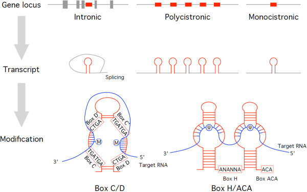
Figure 1.
Secondary structure of snoRNAs and genomic loci. Three types of snoRNA gene loci (top), intermediate transcripts (middle), and mature box C/D and box H/ACA snoRNAs associated with target RNAs (bottom) are shown. Circles indicate modification sites for methylation (m) and pseudouridylation (Ψ). snoRNAs, snoRNA gene loci, and target RNAs are shown in red, gray, and blue, respectively.

