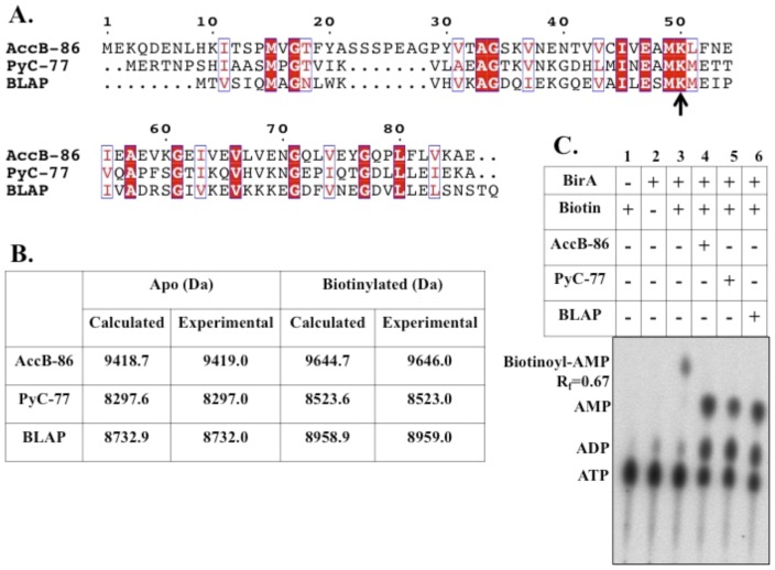
Figure 5. In vitro biotinylation of the B. subtilis biotin acceptor proteins.
A. Sequence alignment of the B. subtilis biotinylated proteins. Conserved residues are in white text and highlighted in red and similar residues are in red text and boxed in blue. The black arrow indicates the conserved lysine residue that becomes biotinylated. B. Mass spectrometry values for purified acceptor proteins AccB-86, PyC-77, and BLAP. C. Thin layer chromatographic analysis of B. subtilis BirA ligase reaction: synthesis of Bio-5′-AMP and transfer of biotin to AccB-86, PyC-77, and BLAP.

