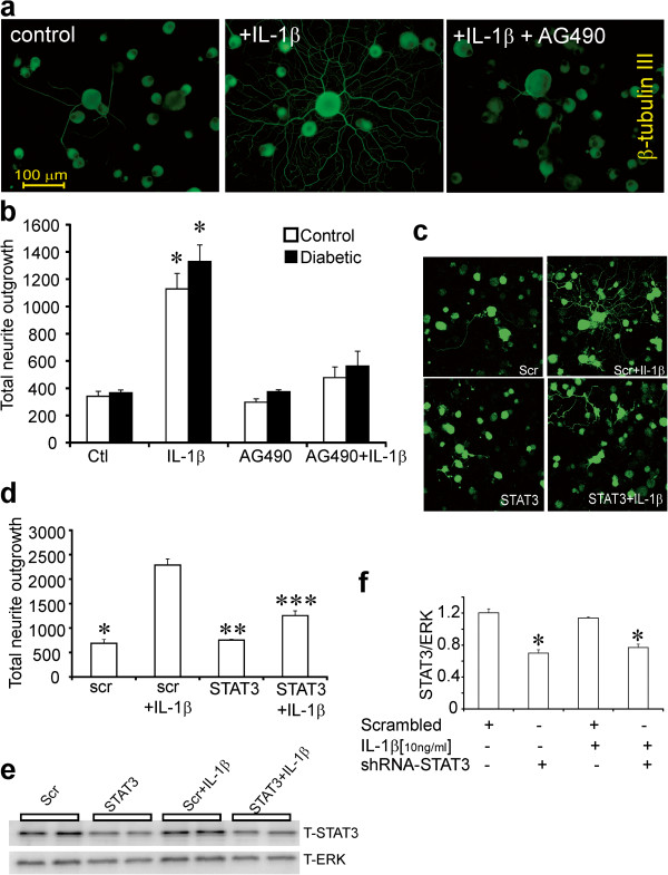
Figure 4.

Blockade of STAT3 signaling inhibits IL-1β-mediated neurite outgrowth. DRG sensory neurons from normal and diabetic rats were cultured for 1 day in the presence of low dose neurotrophic growth factors (to improve viability in presence of AG490) and were exposed to the JAK/STAT inhibitor AG490 (10 μM) ± IL-1β (10 ng/ml). Neurons were fixed and stained for neuron-specific β-tubulin III. Images are shown with (a) no treatment, DRG cultures treated with IL-1β for 24 h or treated with AG490 for 1 h then stimulated with IL-1 β for 24 h. In (b) is shown the impact on total neurite outgrowth in control and diabetic rat cultures. Data are mean ± SEM (n = 3 replicates); *p < 0.05 vs control, or AG490, or AG490+ IL-1β (two-way ANOVA with Bonferroni’s test). In (c and d) is shown total neurite outgrowth levels of DRG cultures from normal rats transfected with plasmids carrying GFP and scramble sequence (scr) or STAT3 shRNA for 48 h and then stimulated with IL-1β (10 ng/ml) for 24 h. Neurite outgrowth was derived from mean pixel area of the GFP signal captured from live cells under confocal microscopy. Data are mean ± SEM (n = 3 replicates); *p<0.05 vs scr + IL-1β, **p<0.05 vs scr + IL-1β, ***p<0.05 vs scr + IL-1β ( oneway ANOVA with Dunnett’s test). In (e) Western blot from DRG cultures studied in (c,d) that were analyzed for T-STAT3 and T-ERK protein. In (f) quantification of expression levels of T-STAT3/T-ERK are expressed and are means ± SEM (n = 4 replicates); *p<0.05 vs scrambled or scrambled + IL-1β ( oneway ANOVA with Dunnett’s test).
