
Figure 5.
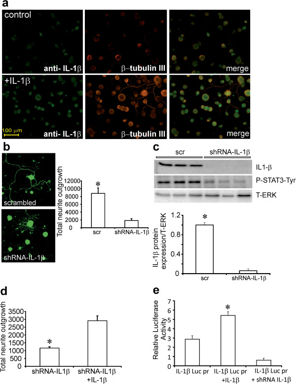
DRG sensory neurons express IL-1β and its blockade inhibits neurite outgrowth. In (a) DRG sensory neurons from normal rats were cultured for 1 day in the presence of low dose of neurotrophic growth factors and were fixed and immunostained for IL-1β. Top panels are untreated cells and bottom panels are treated with IL-1β (10 ng/ml) for 24 h. The right panels are co-stained for β-tubulin III. In (b) DRG cultures were infected with GFP-expressing lentivirus containing scrambled (scr) or shRNA to IL-1β. After 48 h of culture images of GFP fluorescence were acquired and neurite outgrowth quantified. Data are mean ± SEM (n = 3 replicates); *p<0.05 vs shRNA-IL-1β (Student’s t-Test). In addition, in (c) infected cultures were examined by Western blot for IL-1β expression and P-STAT3-Tyr and revealed significantly reduced expression in presence of shRNA to IL-1β. In (d) DRG neurons over-expressed shRNA to IL-1β for 48 h and then were treated ± IL-1β. Neurons were stimulated with/without IL-1β for 24 h and then neurite outgrowth quantified. Data are mean ± SEM (n = 3 replicates); *p < 0.05 vs shRNA for IL-1β + IL-1β (Student’s t-Test). In (e) IL-1β treatment for 24 h up-regulates IL-1β luciferase promoter activity in cultured DRG sensory neurons. Co-transfection with plasmid expressing GFP and carrying shRNA to IL-1β blocked the effect of exogenous IL-1β. Data are mean ± SEM (n = 3 replicates); *p<0.05 vs all groups (oneway ANOVA with Dunnett’s test).