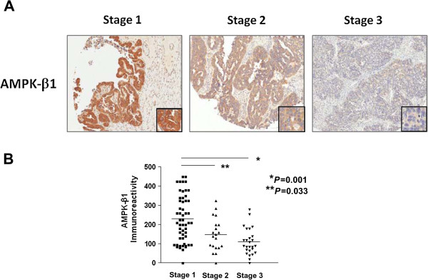
Figure 1.
Expression of AMPK-β1 in ovarian cancer samples. (A) Immunohistochemical analysis of AMPK-β1 expression using an ovarian cancer tissue array (OVC1021, Pantomics). Representative images showing the AMPK-β1 expression in serous subtype ovarian cancer. Tumor stage 1 has the highest level of AMPK-β1, while tumor stage 3 has the lowest level of AMPK-β1 (10x). (B) A graph showing the stepwise decrease of AMPK-β1 expression from stage 1 to stage 3 ovarian cancers.

