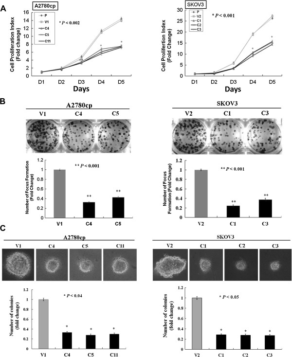
Figure 2.
Overexpression of AMPK-β1 inhibits cell proliferation and anchorage-independent growth ability. (A) XTT cell proliferation assay showing that enforced expression of AMPK-β1 in A2780cp (C4, C5 and C11) (P < 0.002) and SKOV3 (C1, C2 and C3) (P < 0.001) clones displaying a 45 to 50% decrease in the cell growth rate compared with the empty vector (V1) and the parental (P) cell control. (B) Focus formation assay showing that the size and number of foci was reduced 2.5- to 3-fold in AMPK-β1 stable clones of A2780cp (C4 and C5) (P < 0.001) cells and 3- to 4-fold in AMPK-β1 stable clones of SKOV3 (C1 and C3) (P < 0.001) cells compared with the vector controls. (C) Soft agar assay revealing that the AMPK-β1 stable clones of A2780cp (C4, C5 and C11) (P < 0.04) and SKOV3 (C1, C2 and C3) (P < 0.05) cells had a 2.5- to 3-fold reduction in the size and number of colonies compared with the control. P: parental. V, V1 or V2: empty vector controls.

