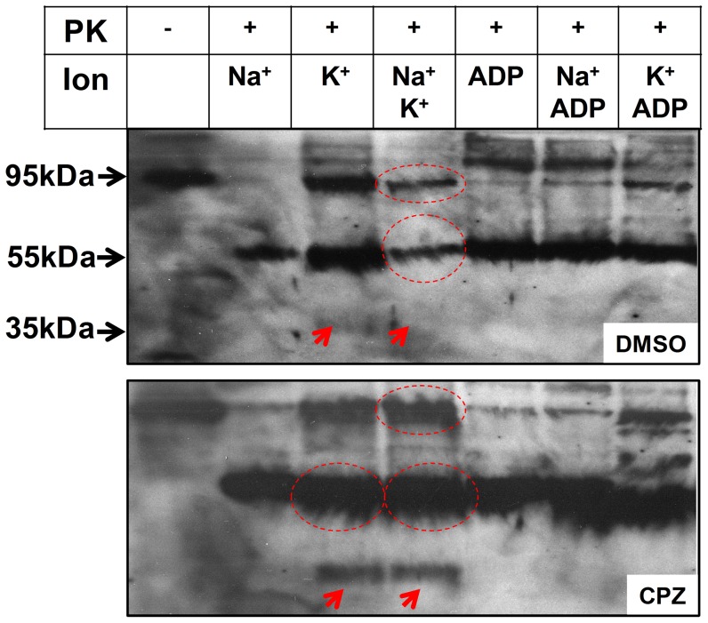
Figure 6. Proteinase K cleavage.
Pig kidney membranes were incubated in 25/or 1 mM ADP, as indicated. The samples were treated with PK (PK/protein ratio ∼1∶50) and the mixture incubated for 40 min at 24°C, and terminated with SDS sample buffer acidified with TCA. Proteolytic products were separated using SDS-PAGE and protein fragments on the gel were transferred to PVDF membranes and visualized by Western blotting using NKA1012-1016 antibody. The labels to the left indicate the apparent molecular weights of the fragments, determined by the use of Precision plus protein standards from Bio-Rad, Cat#161-0363.

