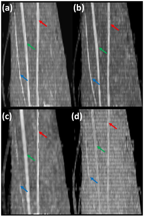
Figure 1.
Oblique sagittal MIP images of FA maps of left forearm acquired with different spatial resolution, different number of diffusion gradient encoding directions (DGED) and repetitions: (a) spatial resolution (1×1 mm2), 42 DGED, and 2 repetitions; (b) spatial resolution (1×1 mm2), 21 DGED, and 4 repetitions; (c) spatial resolution (1.8×1.8 mm2), 42 DGED, and 2 repetitions; (d) spatial resolution (1×1 mm2), 21 DGED, and 1 repetition. The diffusion weighted images are acquired with 8 channel flexible small extremity coil. Median (green arrow), ulnar (red arrow) and superficial radial nerves (blue arrow).

