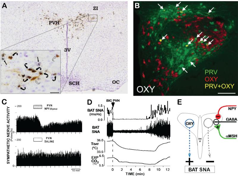
Figure 4. Paraventricular hypothalamic nucleus (PVH) mechanisms influencing BAT thermogenesis.
(A) Histological section through the PVH illustrating the overlap of transynaptically infected, PRV-labeled neurons (brown) following PRV injections into interscapular BAT and in situ hybridization for melanocortin 4-receptor (MC4-R) mRNA expression (black granules). Inset: High magnification of the outlined portion of the PVH. Note the presence of PRV in neurons surrounded by MC4-R (curved black arrows) and PRV in neurons without associated MC4-R (curved open arrows). Bar = 25mm. Modified from (Song et al., 2008).
(B) Immunolabeling of PVH neurons for transynaptic infection with PRV (green) after PRV injections into interscapular BAT and for oxytocin (OXY; red). Arrows indicate neurons containing both PRV and OXY (yellow). Modified from (Oldfield et al., 2002).
(C) Microinjection of neuropeptide Y (NPY), but not saline vehicle, into the PVH inhibited BAT SNA. Modified from (Egawa et al., 1991).
(D) Nanoinjection of bicuculline (BIC) into the PVH completely reversed the cooling-evoked increases in BAT SNA, BAT temperature (TBAT) and expired CO2 (Exp CO2). Modified from (Madden and Morrison, 2009).
(E) Schematic of the proposed neurocircuitry mediating influences on BAT thermogenesis mediated by PVH neurons and their GABAergic regulation. Based in part on (Cowley et al., 1999).

