Figure 6.
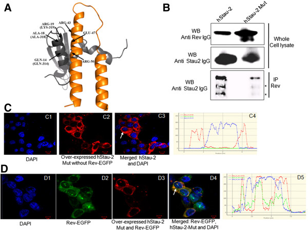
Rev-hStau-2 interactions are essential for hStau-2 dependent promotion of Rev activity. A) The docked complex of Staufen homolog from mouse (1UHZ, grey ribbon) and Rev protein (2X7L, orange ribbon) showing interacting residues: The corresponding residues of hStau-2 protein are indicated in brackets. B) Co-immunoprecipitation of hStau-2 or hStau-2Mut with Rev using anti-Rev antibody: HEK293T cells were transfected with Rev-EGFP vector and co-transfected with hStau2-59 or hStau-2Mut-CMV vector, cell lysates were prepared after 48 hours followed by IP and WB. Whole cell lysates were checked for Rev expression by anti-Rev antibody and for hStau-2 expression by anti-hStau-2 antibody. * indicates non-specific bands or degraded protein. C) Localization of hStau-2Mut. hStau-2Mut localizes in the cytoplasm: HEK293T cells were transfected with hStau-2Mut-CMV vector and stained with goat anti-Staufen-2, Rabbit anti-goat Alexa Fluor 568 antibody and nucleus was stained by DAPI. Distribution plot is shown at the end of the panel. D) Co-localization of Rev and hStau-2Mut: HEK293T cells were transfected with Rev-EGFP and hStau-2Mut-CMV vector. Localization of GFP-tagged Rev was determined by green fluorescence, hStau-2Mut was determined by goat anti-Staufen-2, Rabbit anti-goat Alexa Fluor 568 antibody and nucleus was stained by DAPI. Merged images show co-localization of hStau-2Mut and Rev. Distribution plot is shown at the end of the panel. The precise cells whose graphical analyses have been shown are indicated by the white arrows.

