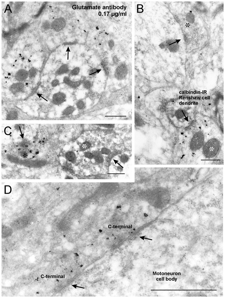
Figure 4. Glutamate-IR is enriched in VAChT-IR motoneuron synapses contacting Renshaw cell dendrites.
Examples obtained with antibody concentration of 0.17 µg/ml. A, Three VAChT-IR boutons surround and make synapses (arrows) on a calbindin-IR Renshaw cell dendrite. Glutamate-IR 10 nm colloidal gold particles are enriched inside VAChT-IR motoneuron terminals compared to surrounding neuropil or calbindin-IR Renshaw dendrites. B, Glutamate-IR colloidal gold particles inside a VAChT-IR motoneuron synaptic bouton (white asterisk) making a synapse (arrow) with a small caliber calbindin-IR Renshaw cell dendrite and an adjacent excitatory synaptic bouton (black asterisk) establishing another synapse (arrow) on a different larger dendrite. Usually VAChT-IR synaptic boutons contain lower densities of glutamate-IR gold particles than adjacent unlabeled terminals. C, Calbindin-IR Renshaw cell synaptic terminal (containing DAB labeling, white asterisk) making a synapse (arrow) with an unlabeled dendrite. These inhibitory terminals contained glutamate-IR at densities similar or below neuropil labeling. A nearby VAChT-IR terminal is making a synaptic contact (arrow) with a dendrite of unknown origin and contains higher levels of glutamate IR gold particles. D, VAChT-IR C-boutons (silver deposits) contacting a motoneuron soma. Significant levels of glutamate -IR colloidal gold particles are found inside the C-boutons and to a lesser extent the motoneuron cell body. These synapses have a characteristic subsynaptic cistern in the postsynaptic site (arrows). Scale bars are 0.5 µm in A, B and C; 1 µm in D.

