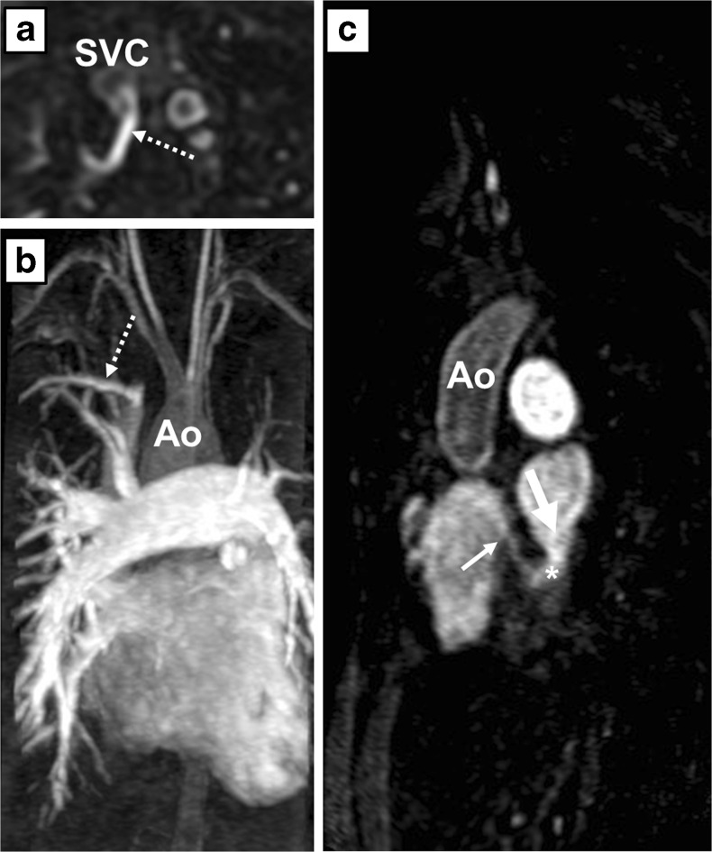
Fig. 2.
Pulmonary angiogram by contrast-enhanced magnetic resonance angiography (MRA) of patient A. Axial (a) and sagittal (c) oblique multiplanar reconstruction and coronal maximum intensity projection (b) of the unroofed coronary sinus (*), the coronary sinus defect (thick arrow) and the entry of the coronary sinus into the right atrium (thin arrow). The right upper lobe drains into the right superior vena cava (dotted arrow). RA right atrium, LA left atrium, Ao aorta, SVC superior vena cava

