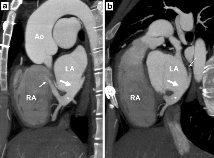
Fig. 3.
Computed tomography angiography (CTA) images after intravenous contrast administration in patient A. a: Sagittal oblique multiplanar reconstruction of the unroofed coronary sinus (*), the coronary sinus defect (thick arrow) and the entry of the coronary sinus into the right atrium (thin arrow). b: Note that the great cardiac vein is not opacified by intravenous contrast (dotted arrow) and that the coronary sinus shows opacification comparable to the left atrium. RA right atrium, LA left atrium, Ao aorta

