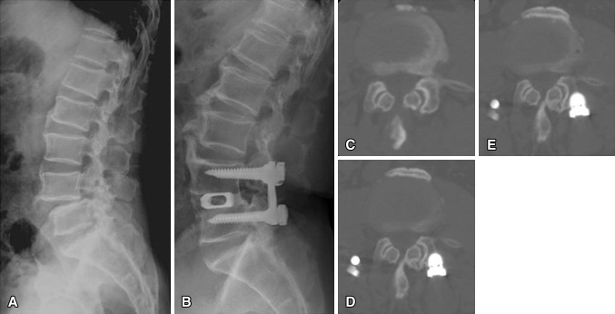
Fig. 4A–E.
Images illustrate the case of a 63-year-old man who underwent anterior lumbar interbody fusion at L4–L5 and experienced radiographic adjacent segment degeneration at followup. (A) A preoperative radiograph shows isthmic spondylolisthesis at L4–L5. (B) A 10-year followup radiograph shows solid fusion and degenerative changes. Compared with the (C) preoperative CT findings, progressive facet degeneration of the cranial segment was evident (D) 5 and (E) 10 years later.

