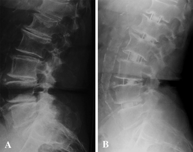
Fig. 3A–B.

(A) Preoperative and (B) 24-month lateral radiographs demonstrate sagittal correction after stand-alone lateral lumbar interbody fusion. Note that, by positioning the cages anteriorly in the disc space, it is possible to increase lordosis, while a posterior positioning of the cage generates a prokyphotic construction.
