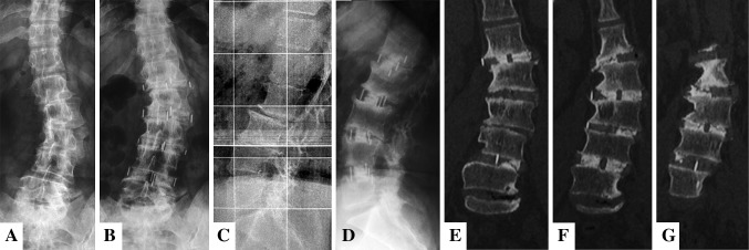
Fig. 4A–G.

(A) Preoperative and (B) 24-month AP radiographs; (C) preoperative and (D) 24-month lateral radiographs; and (E–G) 24-month coronal CT reconstructions demonstrate sagittal and coronal correction and interbody fusion in (E) L1-L2, (F) L2-L3 and L4-L5, and (G) L3-L4. Note also laterolisthesis correction and vertebral derotation.
