
Figure 7.
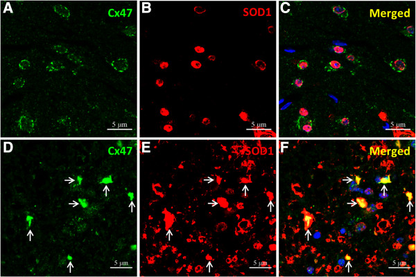
Overexpression of SOD1 in oligodendrocytes of mSOD1-Tg mice. (A-C) Immunoreactivity for SOD1 is observed in the nuclei and cytoplasm of oligodendrocytes in the anterior horns of non-Tg mice at 20 weeks of age and immunoreactivity for Cx47 is preserved at the surface of oligodendrocytes. (D-F) However, SOD1 is markedly accumulated in neurons and astrocytes in the anterior horns of mSOD1-Tg mice at 20 weeks of age and accumulation of SOD1 was also observed in oligodendrocytes with Cx47-immunopositivity in their cytoplasm (arrows). Membranous staining of Cx47 is not visible in abnormal-shaped oligodendrocytes. The nucleus is stained with DAPI (A-F blue). Scale bar; 5 μm (A-F).