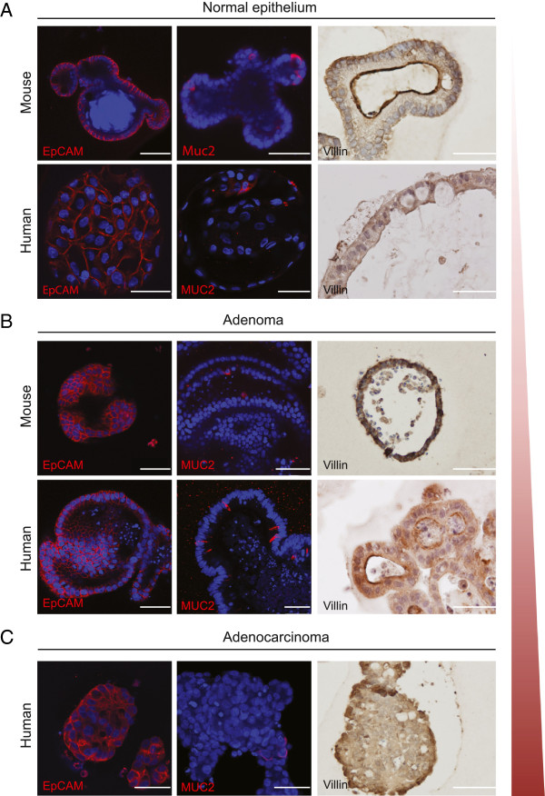
Figure 1.
Multi-lineage potential of intestinal cultures from multiple stages of CRC development. A representative overview of confocal and immunohistochemistry images displaying epithelial cells (EpCAM), goblet cells (MUC2) and enterocytes (Villin) in organoid cultures of (A) healthy epithelial derived from small intestine (mouse) and colon (human), (B) adenoma (C) adenocarcinoma (human). Scale bar: 50 μm.

