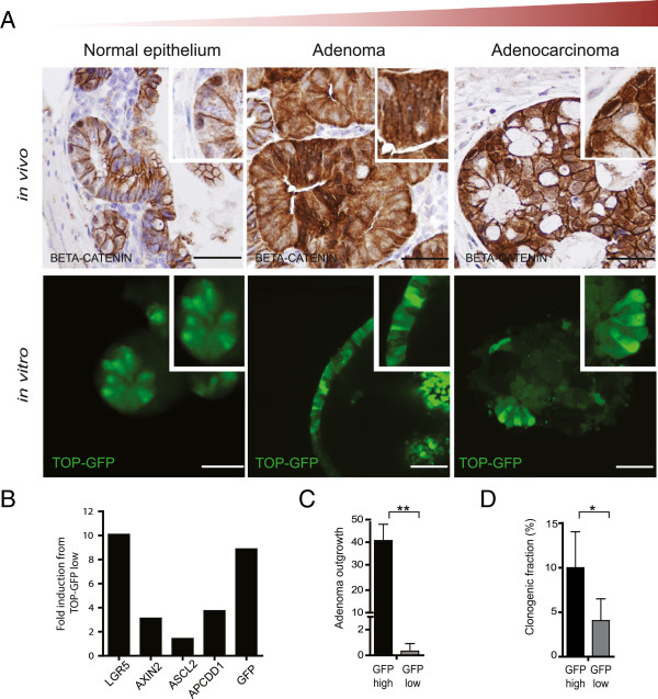
Figure 3.
Functional Wnt activity is maintained throughout CRC development. (A) Immunohistochemistry staining from mouse intestinal epithelial, mouse adenoma, and human adenocarcinoma sections show a heterogeneous β-catenin intracellular distribution indicating Wnt signaling hierarchy in vivo (upper panel). Images are taken at 20× magnification. Lentiviral transduction of TOP-GFP reporter in the cultures derived from the corresponding tissues shows the heterogeneity of Wnt activity in vitro (lower panel). The confocal microscopy images are taken at 63× magnification. (B) Representative graph of qRT-PCR analysis from TOP-GFPhigh and TOP-GFPlow fractions of mouse adenoma cultures. The mRNA values of several Wnt target genes and stem cell markers were first normalized with GAPDH mRNA and expressed as fold induction compared to TOP-GFPlow. (C and D) Colony-forming efficiency in the sorted TOP-GFP cells showing that TOP-GFPhigh cells exhibit higher clonogenic potential in both mouse (C) and human (D) adenoma cultures. Data shown represents mean ± SD from three independent experiments from mouse adenoma cultures (N=2) and human adenoma cultures (N=2). Scale bar, 50 μm. * p<0.05, ** p<0.01, ***p<0.0001 (t-test).

