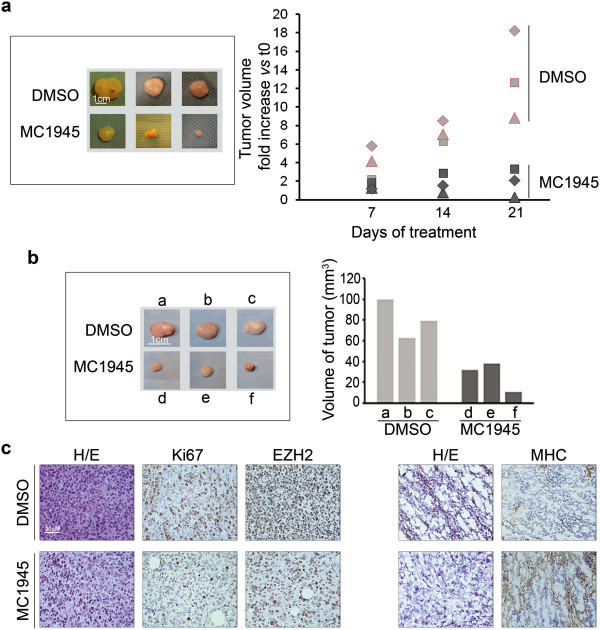
Figure 7.
Analysis of pharmacological inhibition of EZH2 in vivo. RD cell suspensions in PBS (10×106 cells) were injected subcutaneously into the posterior flanks of athymic 6-week-old female BALB/c nude mice (nu + \nu+). (a) As soon as the tumors became palpable, i.e., about 2 months after the initial inoculation when they reached approximately 70–80 mm3, mice were intraperitoneally injected with MC1945 (2.5 mg/Kg) or control vehicle (DMSO) twice daily, 3 days per week for 3 weeks when mice were sacrificed with no visible signs of toxicity. Tumor volume was measured by caliper weekly for 21 days (right) after which xenografts were surgically removed (left). (b) for immunohistochemical studies, a group of mice were sacrificed at day 12 of treatment, during the exponential tumor growth phase, and xenografts excised. Tumor volume was measured by caliper prior to the initiation of treatment and at the time of sacrifice. (b, left) xenografts from either nude mice treated with MC1945 (d, e, f) or with DMSO (a, b, c) as controls. (b, right) Portions of the excised tumors in (b) were embedded in paraffin for immunohistochemical analysis or snap-frozen in liquid N2. Histogram reports tumor volumes of each xenograft. (c) Sections of 10 μm cut from xenograft blocks were stained with hematoxylin/eosin. Five μm serial sections were subjected to immunohistochemistry for the expression of EZH2 and Ki67 (paraffin-embedded, nuclear orange staining in left panels) and MHC (OCT embedded, staining with the MF-20 antibody, cytoplasmic orange staining in right panels) of serial DMSO (upper panels) and MC1945 treated (lower panels) sections from RD xenografts. Counterstaining was carried out with Gill’s hematoxyline (Bio-Optica, MI, Italy). Sections were dehydrated and mounted in mounting medium. Images were acquired under an Eclipse E600 microscope (Nikon) through the LUCIA software, version 4.81 (Nikon) with a Nikon Digital Camera DXM1200F.

