Figure 3.
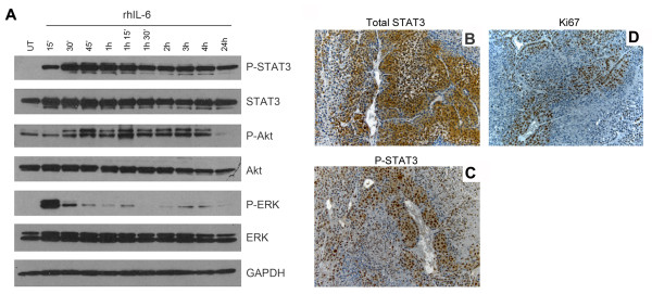
STAT3 phosphorylation in xenograft human oral squamous cell carcinomas correlates with tumor cell proliferation and presence of blood vessels. A, HeLa cells were serum-starved overnight and exposed to 20 ng/ml rhIL-6 for the indicated time points. Phosphorylated and total levels of STAT3, Akt, and ERK were determined by Western blots. B-D, xenograft human tumors were generated in SCID mice by co-implanting HeLa and HDMEC. Tumors were retrieved after 28 days, and tissues were analyzed by immunohistochemistry: B, total STAT3 with cytoplasmic localization, diffused through the tissue; C, phosphorylated STAT3 with nuclear localization, concentrated in the proximity of blood vessels; D, Ki67 with nuclear translocation, localized primarily around blood vessels. Photomicrographs at 200×.

