
Figure 1.
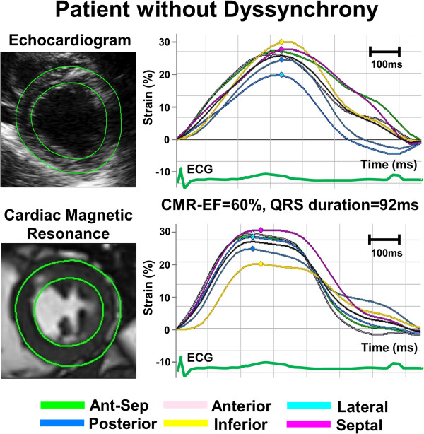
Example of radial time-strain curves by speckle tracking echocardiography (top panel) and feature tracking CMR (bottom panel) in a patient with normal left ventricular (LV) function and without dyssynchrony, demonstrating synchronous time-to-peak-strain curves.