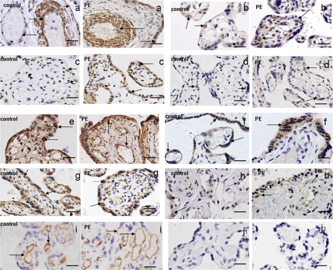
Figure 1.
A-H, The expression profile of Notch receptors 1-4 (A-D) and its ligands Jag1 and 2 (E and F) and Dll1 and 4 (G and H) in placentas from patients with early-onset severe preeclampsia, by immunohistochemical staining. Notch1 was predominantly expressed in the endothelial cells and the vessel walls of the placental villi (A). In addition, there was weak Notch-1 immunopositivity in the cytoplasm of syncytiotrophoblasts (A). Notch 2, 3, and 4 immunopositive cells were mainly cytotrophoblast cells (B-D). Jag1 was detected in cytotrophoblast cells as well as in the endothelial cells of the placental villi (E). Jag2 was mainly found in the cytoplasm of syncytiotrophoblasts (F). Dll1 and Dll4 were predominantly immunopositive in the cytotrophoblast cells (G, H: solid arrows) together with a faint immunopositivity for Dll4 in the endothelial cells (H: dashed line arrows).The positive controls (anti-CD34 staining) was displayed in (I), and the negative control (isotype-matched control) was shown in (J1; Mouse IgG2B) and (J2; Rabbit IgG). Scale bar: 20 μm.

