Figure 1.

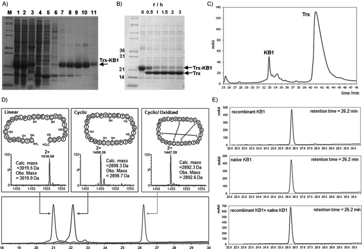
Production of KB1. A) SDS-PAGE analysis of Ni2+-affinity-purified Trx-KB1 fusion protein. M: molecular weight markers. Lane 1: whole-cell lysate, lane 2: soluble fraction, lane 3: insoluble fraction, lane 4: column flow-through, lane 5: column wash (5 mm imidazole), lane 6: column wash (20 mm imidazole), lanes 7–11: eluted fractions (40–500 mm imidazole). B) TEV protease digestion of the fusion protein shows accumulation of Trx (released linear KB1 is not visible on the gel). C) Preparative HPLC allows straightforward separation of the released KB1 from Trx. D) Analytical HPLC (lower panel) and MS (upper panels) characterization of purified linear, cyclic (reduced) and folded KB1 samples. E) HPLC coelution experiment: KB1 (upper panel), native KB1 (isolated from O. affinis, middle panel) and a 1:1 mixture of each peptide (lower panel).
