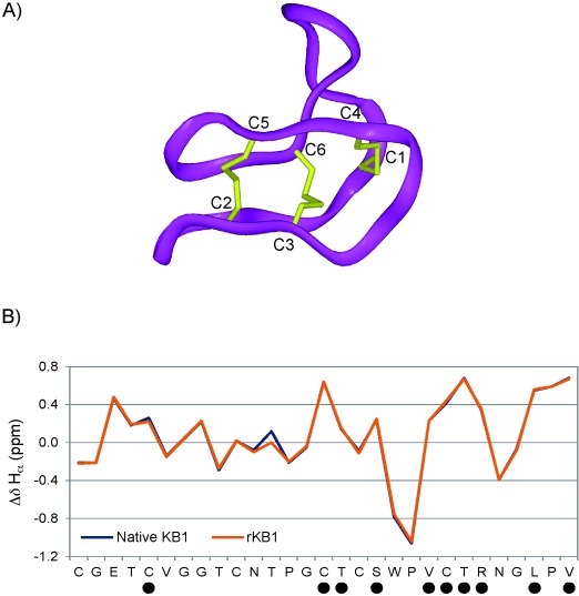
Figure 2.

Characterisation of kalata B1 through NMR spectroscopy. A) A ribbon representation of the native KB1 backbone structure, including disulfide-bonded cysteines (in yellow). B) A graphical depiction of the deviation in Hα chemical shift from random coil values for each residue in native KB1 (blue) and our semisynthetic KB1 (orange). • denotes residues in which slow-exchanging amide protons have been identified (Figure S7).
