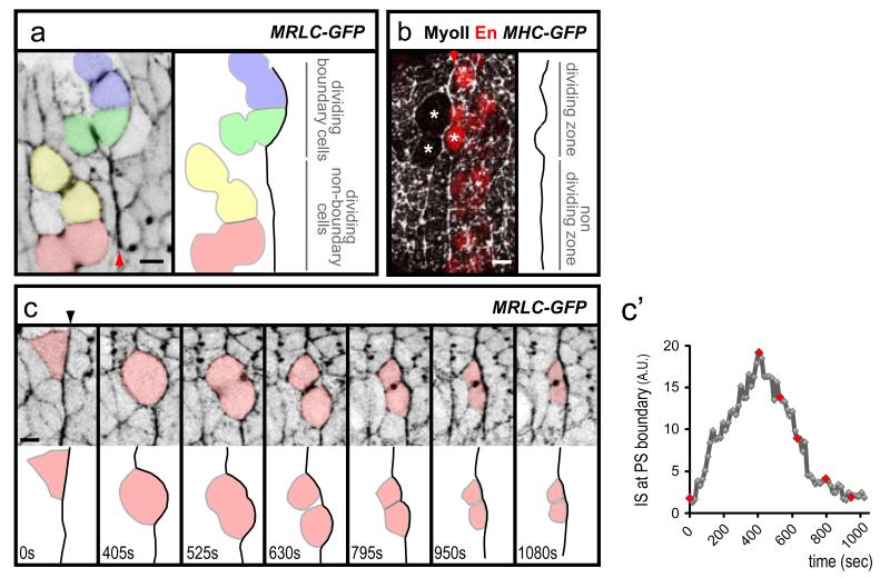
Fig. 3. Cell divisions challenge PS boundaries.
(a) Movie frame showing that dividing boundary cells (coloured blue and green), but not non-boundary cells (red and yellow) deform the MyoII cable at a PS boundary (identified by MRLCGFP enrichment, arrowhead). (b) The MyoII cable (arrowhead) is not dismantled when boundary cells divide (stars). (c) Movie frames of a MRLC-GFP embryo showing how the division of a boundary cell deforms transiently the MyoII cable (identified by MRLCGFP enrichment). c’) Quantification of membrane straightness for the first 6 frames in c (red dots). Note that, in contrast to all other quantifications which consider several boundary cells, the IS measured here corresponds to the length of the interface of the boundary cell coloured in red.

