Figure 1.

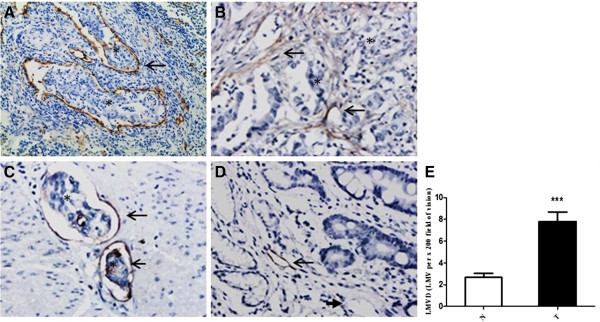
Immunohistochemical labeling of lymphatic microvessels (EnVision™) and LMVD in primary gastric cancer tissue and noncancerous gastric tissue. (A-D) Lymphatic microvessels labeled with D2-40 by IHC (EnVision™). (A, B) Gastric cancer (×100), (A) peritumoral lymphatic vessels were large, within which was nests of no staining tumor cells (asterisks); (B) intratumoral lymphatic vessels had narrow and collapsed lumen, with irregular cell walls (long arrow). (C) Invaded muscle tissue (×200). There were a few tumor cells in dilated lymphatic microvessels, as ‘lymphatic vessel invasion’ (asterisks). (D) Non-cancerous mucosa (×200). Long arrows indicated lymphatic microvessels, and short arrows denoted blood capillaries. Asterisks represented the areas where tumor cells are. (E) LMVD in gastric cancer tissue (T) was statistically higher than that in the non-cancerous counterparts (N) (*** P <0.001).
