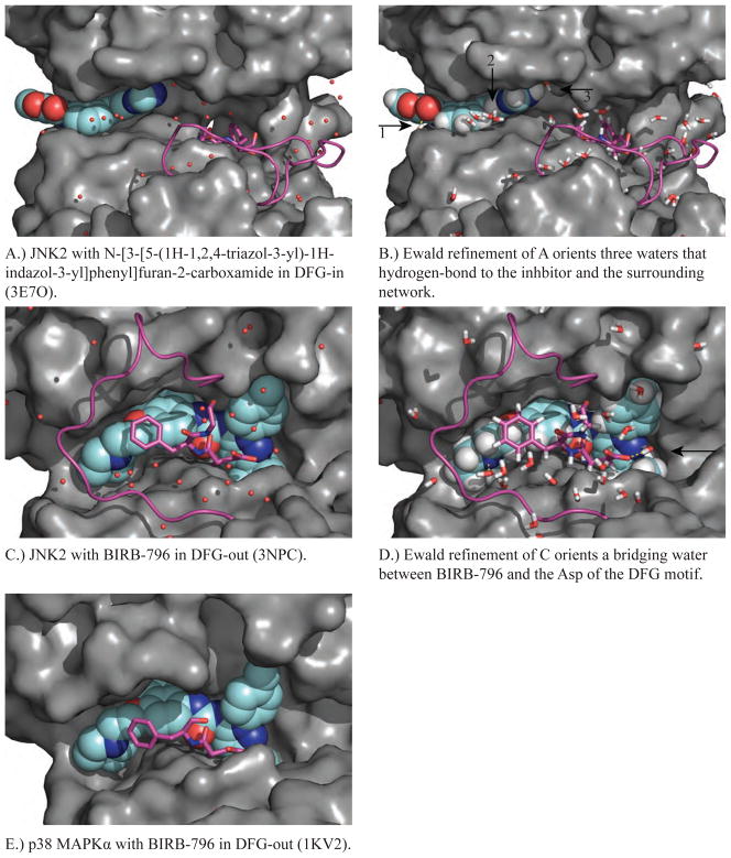
Figure 4.
Shown in each panel is a MAP kinase structure complexed with an inhibitor (cyan, spacefill) that targets DFG-in or DFG-out (magenta, ball & stick) and the corresponding conformation of the activation loop (magenta, backbone only). A.) JNK2 in the DFG-in conformation is shown in a complex with type-I inhibitor N-[3-[5-(1H 1,2,4-triazol-3-yl)-1H-indazol-3-yl]phenyl]furan-2-carboxamide (PDB ID 3E7O). B.) Ewald refinement of A orients the water hydrogen-bonding network around the JNK2 inhibitor-binding site. C.) JNK2 in the DFG-out conformation in a complex with type-II inhibitor BIRB-796 (PDB ID: 3NPC). D.) Ewald refinement of C orients the water hydrogen-bonding network around the JNK2 inhibitor-binding site. E.) p38 MAPKα in the DFG-out conformation in a complex with BIRB-796 (PDB ID 1KV2). Ewald refinement was not performed for E because no diffraction data was deposited.

