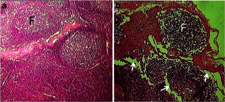
Fig. 4.
The transverse sections of the sub-iliac lymph node in the control (a) and 180 mg/kg phenol-treated (b) animals. The b section shows significant decrease in the size of the follicles (F) as well as empty spaces (arrows) around the follicles in the phenol treated mice (hematoxylin and eosin stain; a, b ×400)

