Figure 2.

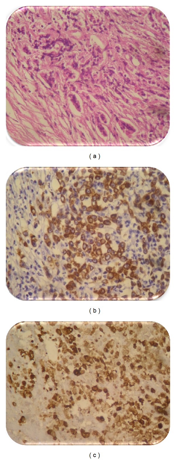
(a) Histopathological findings showed cells difficult to typify (40). (b) Immunohistochemistry of the piece showed intense expression of cytokeratin (AE1/AE3). (c) Expression of CK20 similar to the primary colorectal carcinoma.

(a) Histopathological findings showed cells difficult to typify (40). (b) Immunohistochemistry of the piece showed intense expression of cytokeratin (AE1/AE3). (c) Expression of CK20 similar to the primary colorectal carcinoma.