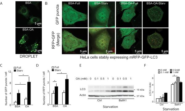
Figure 1. Preformed lipid droplets enhance starvation-induced autophagy.
(A) and (B) HeLa cells stably expressing mRFP-GFP-LC3 were treated for 20 h with BSA alone (BSA) or with BSA-oleic acid (OA; 500 μM OA) and starved (Starv) in EBSS for 90 min or incubated in full medium (Full). (A) Visualization of lipid droplet accumulation. Lipid droplets were stained with Bodipy 493/503. (B) Confocal images of samples (for images in high content acquisition mode see Figure S1). (C) and (D) Number of GFP+ puncta (C) and RFP+ puncta (D) per cell were quantified by high content image acquisition and analysis. Data: means ± s.e. (n=3, where n represents separate experiments; each experimental point in separate experiments contained >500 cells identified by the program as valid primary objects); *, p<0.05 (t-test). (E,F) HeLa cells treated with OA as in A–D with or without bafilomycin A1 (BafA1); LC3-II/actin ratios determined by immunoblotting (E) and densitometry (F). Immunobloting data: means ± s.e., *, p<0.05 (t-test).

