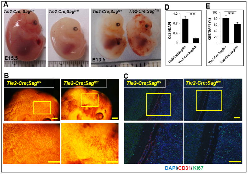
Figure 1. Sag endothelial deletion disrupts vascular development in mouse embryos.

(A) Appearance of mouse embryos at E15.5 (left) and E13.5 (right). (B) Appearance of the brain regions of mouse embryos at E13.5 after CD31 whole-mount staining. Scale bar represents 3 mm. (C) Sagittal sections of control and mutant embryos were stained with antibodies against CD31, Ki67, and DAPI at the spinal cord areas. Scale bar represents 100 μm. (D and E) Quantification of CD31 positive cells (D) and Ki67 positive cells (E). The data were normalized as the ratio of CD31+/DAPI+ cells and represented as fold change with CD31+/DAPI+ value setting at 1 in Tie2-Cre;Sagfl/+ embryos (mean ± SEM, n=3) (D). The number of cells expressing Ki67 was normalized to the total number of DAPI positive cells as a percentage (mean ± SEM, n=3) (E).
