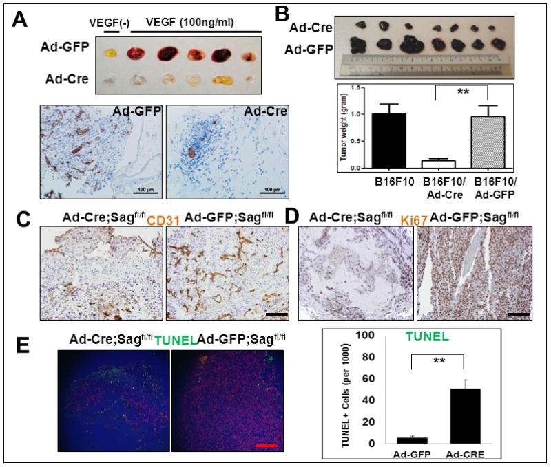
Figure 5. Sag EC deletion inhibits in vivo angiogenesis and tumorigenesis.

Sagfl/fl mice were injected with matrigel mixed with Ad-Cre (on the left flank) or Ad-GFP control (on the right flank) in the presence or absence of VEGF. After 7 days, mice were euthanized and the matrigel plugs were harvested and photographed (A, top panel), then fixed, sectioned and stained with CD31 antibody (A, bottom panels). Sagfl/fl mice were injected with B16F10 mouse melanoma cells (5×10⁵) mixed with Matrigel and Ad-Cre or Ad-GFP as indicated (right before injection without pre-virus infections). Twelve days later, tumors were harvested and photographed (B, left panel). Shown is mean ± SEM from 7 mice in each group, except B16F10 only control group (n=3) (B, right panel). The tumor tissues were fixed, sectioned and stained for CD31 (for blood vessels, C), Ki67 (for proliferation, D) and TUNEL assay (for apoptosis, E). Scale bar represents 100 μm.
