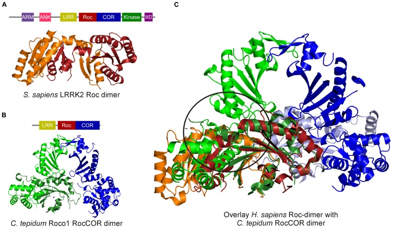
FIGURE 3.
Crystal structures of the human swapped Roc dimer and the C. tepidum RocCOR dimer. (A) Human Roc Dimer depicted as a cartoon with Roc-A in orange and Roc-B in red. Above the domain representation of LRRK2 is shown. (B) Domain representation of C. tepidum and below a cartoon representation of the C. tepidum RocCOR structure with RocCOR-A in green and COR-B in blue. (C) Overlay of the two structures Roc-A (orange) of the human protein clashes with the N-terminal part of the C. tepidum COR-A (green). [PDB: 2ZEJ (human Roc), 3DPU (C. tepidum RocCOR)].

