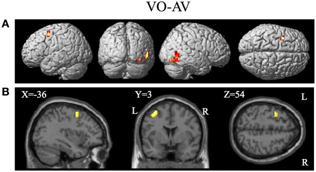
Figure 8.

Brain activity significantly active for the contrast of visual only VO relative to the combined AV conditions thresholded at p < 0.001 uncorrected. Activity was present in the left PMvs/PMd and the left MT/V5 visual motion processing area. (A) Activity rendered on the surface of the left, back, right, and top of the brain. (B) Section through brain taken at MNI coordinate −36, 3, 54 shows activity that was present in the PMvs/PMd region. L, left side of brain; R, right side of brain.
