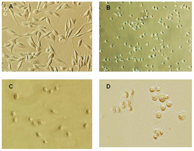
Figure 1. Phase contrast images of MDA-MB-435 cells and isolated nuclei.
(A) MDA-MB-435 cells. (B) Nuclei isolated in isotonic buffer A. Cytoplasm of the cell at this stage was removed by detergent-containing buffer; approximately 65% of proteins were extracted to the cytosol; nuclei contain perinuclear region proteins; nucleoli are visible. (C) Nuclei after extraction of the perinuclear region with buffer B. The core nucleus does not collapse after removal of the perinuclear region proteins; approximately 20% of total cellular proteins were extracted with the perinuclear fractionation; nucleoli are visible. (D) Nuclei isolated with the classical method of using hypotonic buffer. The shape of nuclei varies; some “fibrous” structures around nuclei are visible; nuclei contain nucleoli.

