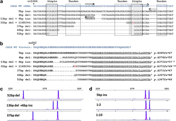
Figure 2.
Characterization of CALR exon 9 mutations in patients with thrombocytosis. (a) Comparative sequences of the seven different types of CALR mutations found in this study against the wild-type CALR exon 9 cDNA. Repeat elements are highlighted by gray solid lined (simple) or dashed lined (tandem) boxes and inverted sequences are depicted by reverse arrows. Presumed genomic regions from which these inversions may have derived are depicted by matching solid/dashed forward arrows. A polymorphism (G) is depicted in red and this was also found in normal DNA from the same individual. (b) Predicted C-terminal amino-acid sequences resulting from the frameshift indel mutations compared with the wild-type calreticulin protein sequence. The common novel peptide sequence shared by all mutations described to date is underlined. The amino-acid variant resulting from the polymorphism above is shown in red (e). (c) Results of PCR fragment analysis of CALR exon 9 in three patients with different indel mutations. (d) Example of fragment analysis from a patient with a 5-bp insertion diluted into normal control DNA shows that the mutation is readily detectable after a 1:10 dilution. †Novel mutation variant, ††Novel mutant amino-acid sequence.

