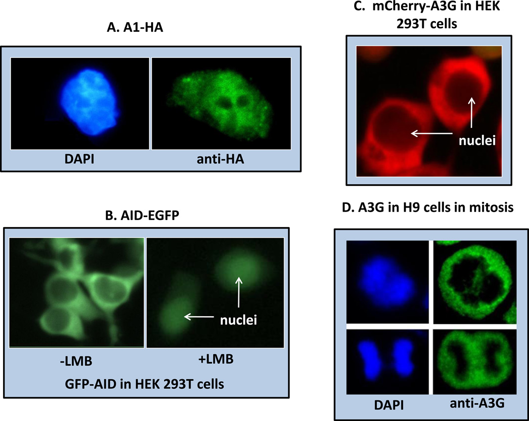
Figure 3. Localization of the APOBEC proteins by in situ fluorescence.
A. APOBEC-1 (A1) with a C-terminal HA tag was expressed by transfection in McArdle rat hepatoma cells. Immunocytochemical localization of the HA tag in fixed cells shows that A1 is distributed throughout the cytoplasm and nucleus (DAPI) but is not localized in the nucleolus. A1 shuttles between the cytoplasm and nucleus. B. Activation Induced Deaminase (AID) as a C-terminal GFP chimeric protein was expressed in transfected human embryonic kidney cells 293T and visualized in live cells. AID appears predominantly in the cytoplasm (left panel, four transfected cells shown) but its rapid shuttling activity can be demonstrated by inhibiting its CRM1-dependent nuclear export with Leptomycin B (LMB) (right panel, two transfected cells shown). C. A3G as an N-terminal mCherry chimeric protein was expressed in transfected HEK293T cells and visualized in live cells. A3G is restricted to the cytoplasm because it has no nuclear localization signal but does have a cytoplasmic retention signal. D. Natively expressed A3G in synchronized H9 cells after fixing and staining with anti-A3G polyclonal antibody. DAPI stained mitotic chromosomes (left top, anaphase; left bottom, telophase) are segregated in the mitotic cells from cytoplasmic A3G. Original magnification was 40X.

