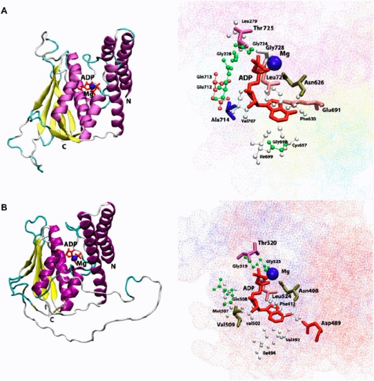
Figure 4.

Cartoon view diagram of the secondary structure of TD domain showing the arrangement of 7 β-sheets and 7 α-helices in AtHK1 (A) and of 5 β-sheets and 7 α-helices in OsHK3b (B) with ADP and Mg along with the marked residues which were observed to form hydrogen bond with ADP. The residues are numbered according to their respective position in the complete sequence of AtHK1 and OsHK3b.
