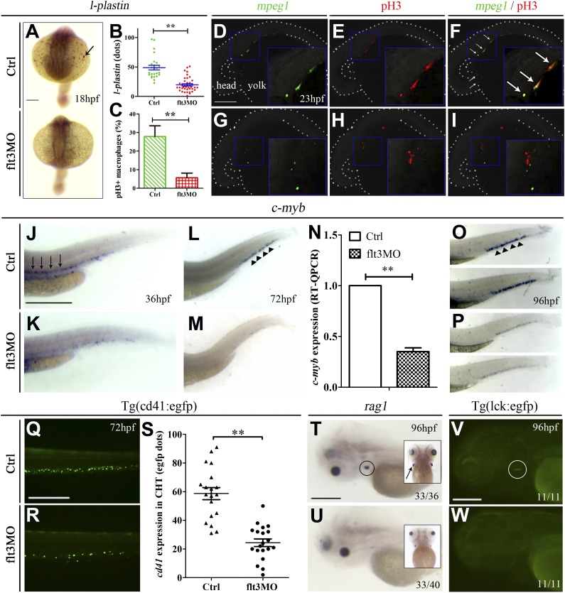
Figure 2.
flt3 knockdown affected primitive myelopoiesis and definitive hematopoiesis. MO targeting zebrafish flt3 was microinjected into 1-cell–stage embryos (6 ng per embryo), and uninjected embryos from the same batch were used as control. (A) WISH comparing the expression of l-plastin between control (top panel) and flt3MO embryos (bottom panel) at 18 hpf. The arrow indicated l-plastin expression at the anterior yolk sac region. (B) Comparison of l-plastin expression (dots in the anterior yolk sac) in control and flt3MO embryos at 18 hpf (corresponding to panel A). (C) Comparison of percentage of Phospho-Histone H3 (pH3) positive macrophages in control and flt3MO embryos. The percentage was calculated based on the number of overlapped signals divided by the number of macrophages (GFP+) in each embryo. The results represented mean ± 1 standard error of the mean of 15 embryos examined in 3 different experiments. (D-I) Phospho-Histone H3 (Ser10) immunostaining showing the proliferation of macrophages (mpeg1:egfp transgenic fish) in control embryos (D-F) and flt3 morphants (G-I) at 22 hpf. The images in panels D-F were obtained from the same embryo, showing the overlapping signals in panel F. It was similarly presented for panels G-I. The body including the developing eyes and yolk sac was outlined by the dotted lines. Proliferative cells (nonmacrophage) were also identified in the heads and tail regions and were shown in supplemental Figure 7G,K. (J-P) WISH showing the c-myb expression in control (J,L,O) and flt3MO (K,M,P) embryos at 36, 72, and 96 hpf, respectively. The arrows in panel J and arrowheads in panels L and O indicated c-myb expression in the ventral wall of DA and CHT. (N) Real-time quantitative PCR comparing c-myb expression between control and flt3MO embryos (corresponding to panels L-M). (Q-S) Comparison of cd41:egfp expression in CHT between control (Q) and flt3MO (R) embryos using Tg(cd41:egfp) transgenic embryos. (S) Comparison of cd41:egfp expression (dots of egfp+ cells in the CHT) in control and flt3MO embryos (corresponding to panels Q-R). (T-U) WISH showing rag1 expression in control (T) and flt3MO (U) embryos. The circle in panel T and arrow in the inset indicated rag1 expression in the thymus at lateral and ventral views. (V-W) Comparison of lck expression in thymus between control (V) and flt3MO (W) embryos using Tg(lck:egfp) transgenic embryos. The circle in panel V indicated lck expression in the thymus. Scale bars represent 500 μm.

