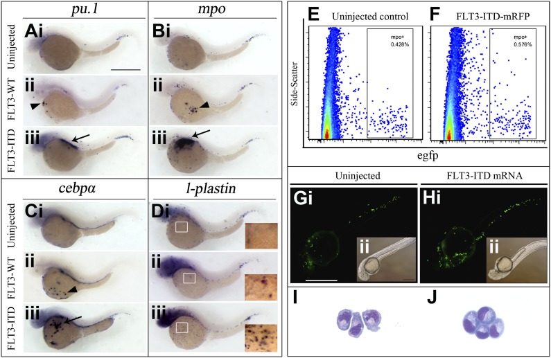
Figure 5.
FLT3-ITD overexpression induced ectopic myeloid expansion in zebrafish. (A-D) Human WT FLT3 (FLT3-WT) and FLT3-ITD mutation (FLT3-ITD) was cloned into pegfp-N3 vector. The vector containing the FLT3WT-T2a-egfp and FLT3ITD-T2a-egfp transgene was microinjected into 1-cell–stage zebrafish embryos. WISH comparing the pu.1 (A), mpo (B), cebpα (C), and l-plastin (D) expression between uninjected, FLT3-WT, and FLT3-ITD overexpressing embryos at 36 hpf. The arrowheads and arrows indicated the typical intermediate and severe expansion (for definition, see supplemental Figure 11). Panel Ai-iii represents the pu.1 expression in uninjected (Ai), FLT3-WT (ii), and FLT3-ITD (iii) embryos, respectively. Panel Bi-iii represents the mpo expression in uninjected (Bi), FLT3-WT (ii), and FLT3-ITD (iii) embryos, respectively. Panel Ci-iii represents the cebpα expression in uninjected (Ci), FLT3-WT (ii), and FLT3-ITD (iii) embryos, respectively. Panel Di-iii represents the l-plastin expression in uninjected (Di), FLT3-WT (ii), and FLT3-ITD (iii) embryos, respectively. (E-F) Human FLT3-ITD was cloned into the pDsRed-Monomer-N1 vector to generate the FLT3-ITD-T2a-mRFP transgene. The CMV-driven FLT3-ITD-T2a-mRFP transgene was microinjected into 1-cell–stage embryos. The level of increase in mpo+ cells might appear lower as the FLT3-ITD-T2a-mRFP transgene expressed weaker FLT3-ITD than the FLT3ITD-T2a-egfp transgene used for WISH. (G-H) Human FLT3-ITD mRNA was in vitro transcribed and microinjected into 1-cell–stage Tg(mpo:egfp) embryos. The egfp+ cells indicate the mpo expression in uninjected (Gi-ii) and FLT3-ITD mRNA (Hi-ii) embryos at 36 hpf. (I-J) FACS of mpo+ cells by egfp, and the morphology of the mpo+ cells in uninjected (I) and FLT3-ITD mRNA (J) injected embryos were examined (Shandon Cytospin 4; Thermo Electron Corporation) with Wright-Giemsa staining. The mpo:egfp+ cells were sorted (MoFlo XDP; Beckman Coulter) Scale bars represent 500 μm in panels A-D, and ×600 magnification in panels I-J.

