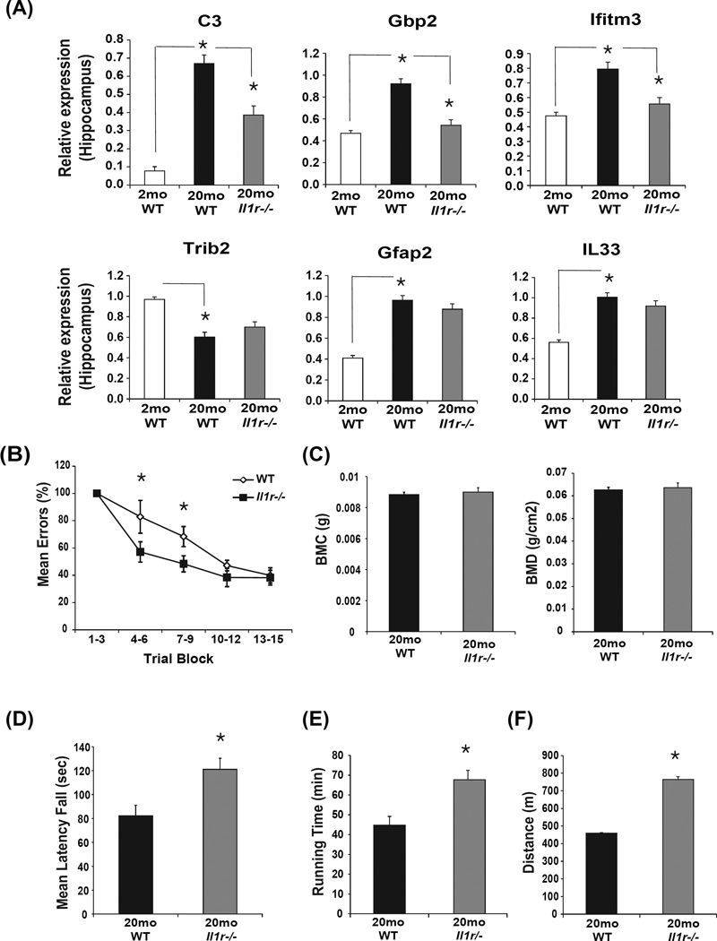
Figure 6. Ablation of IL-1 signaling partially protects against age-related functional decline.
(A) The hippocampi from separate cohort of young (3 month), old (20 month) WT and Il1r−/− mice (n= 6–12/group) were analyzed for complement, interferon and Nlrp3-dependent inflammatory genes using realtime-PCR. (B) The Stone T-maze test in 20-month old WT mice and age-matched Il1r−/− mice (n = 12/group). Mice were given 15 trials in the T-maze with each trial having a maximum length of 300 sec and number of errors during each trial block was recorded. (C) Bone mineral content of femorae of 20-month old WT and Il1r−/− mice (n=12) is measured in grams of calcium hydroxypatite; bone mineral density represents the mineral in bone per area i.e. areal bone mineral density. (D) The mean latency to fall from a rotating rod (Rotarod test) in 20-month old WT and Il1r−/− mice (n=12). (E, F) The treadmill test showing total running time and total distance run by 20-month old WT and Il1r−/− mice (n =12). All data are presented as Mean (SEM)* P<0.05.

