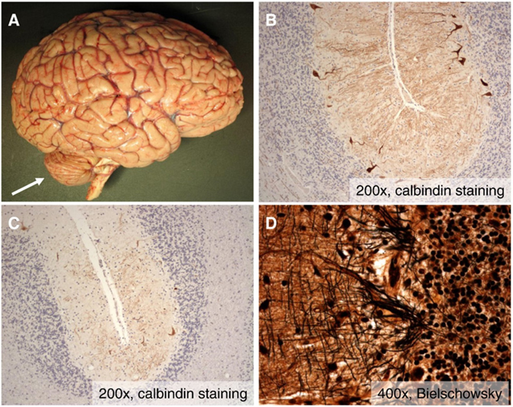
Figure 2.
Cerebellar pathology in variant AT. A) Gross photograph of fresh brain reveals marked cerebellar atrophy (arrow) with no evidence of cortical atrophy or other abnormalities. B–C) Photomicrographs at intermediate magnification (200×) of calbindin immunohistochemistry (which specifically labels Purkinje neurons) demonstrate moderate loss of Purkinje neurons in some areas (B) and severe loss in others (C). D) High magnification photomicrographs of Bielschowsky silver stained sections of cerebellar cortex reveals the presence of scattered “empty baskets” indicating Purkinje neuron dropout.

