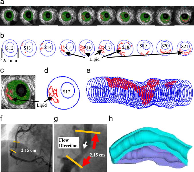
Fig. 3.

IVUS model construction process: (a) selected 10 slices from a 44-slice IVUS data set with IVUS-VH; (b) contour plots; (c) enlarged view; (d) enlarged contour plot; (e) 3D plaque geometry showing lipid core locations; (f) angiography showing location of the lesion and vessel curvature; (g) enlarged angiography showing the lesion; (h) illustration of vessel bending.
