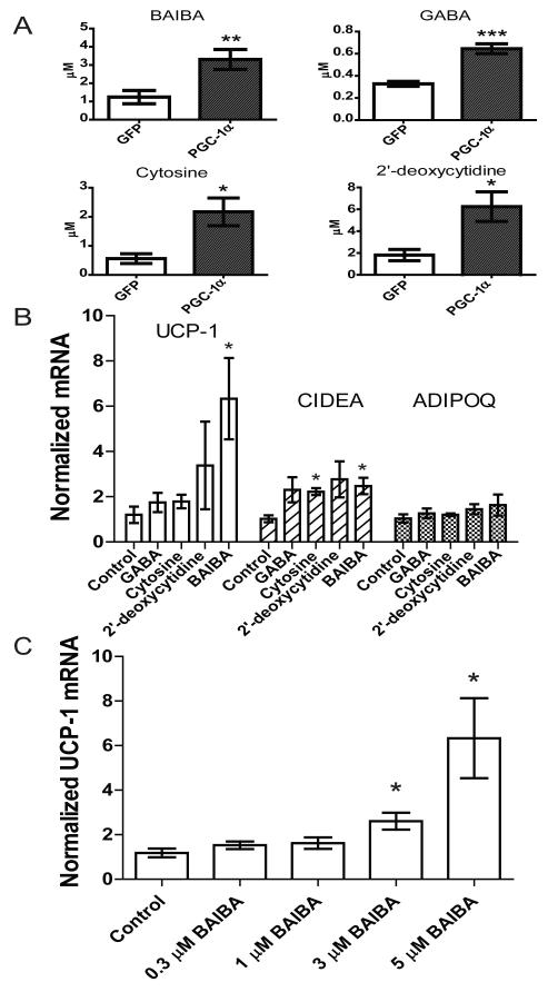
Figure 1. Metabolites accumulate in the media of myocytes as a result of forced PGC-1α expression and stimulate expression of brown adipocyte-specific genes in adipocytes.
A) Myocytes were transduced with an adenoviral vector expressing either PGC-1α (n=6) or GFP (n=6). After 24 hours of exposure to these cells, media was analyzed using an LC-MS based metabolite profiling method measuring 100 small molecules (see Methods). B) BAIBA (5 μM) induces expression of brown adipocyte-specific genes in primary adipocytes differentiated from the stromal vascular fraction isolated from inguinal WAT over 6 days. Additional metabolites tested at physiologically relevant doses included GABA (3 μM), cytosine (1 μM), and 2-deoxycytidine (15 μM). While BAIBA significantly increased the expression of the brown adipocyte-specific genes UCP-1 and CIDEA, it did not alter the expression of the white adipocyte gene adiponectin (ADIPOQ). Cumulative data from a total of 5 independent observations are shown. C) BAIBA concentrations in the low micromolar range significantly and dose-dependently increased the expression of the brown adipocyte-specific gene UCP-1. *, P ≤ 0.05, **, P ≤ 0.01, ***, P ≤ 0.001. Data are represented as Mean ± SEM.

